Case History:
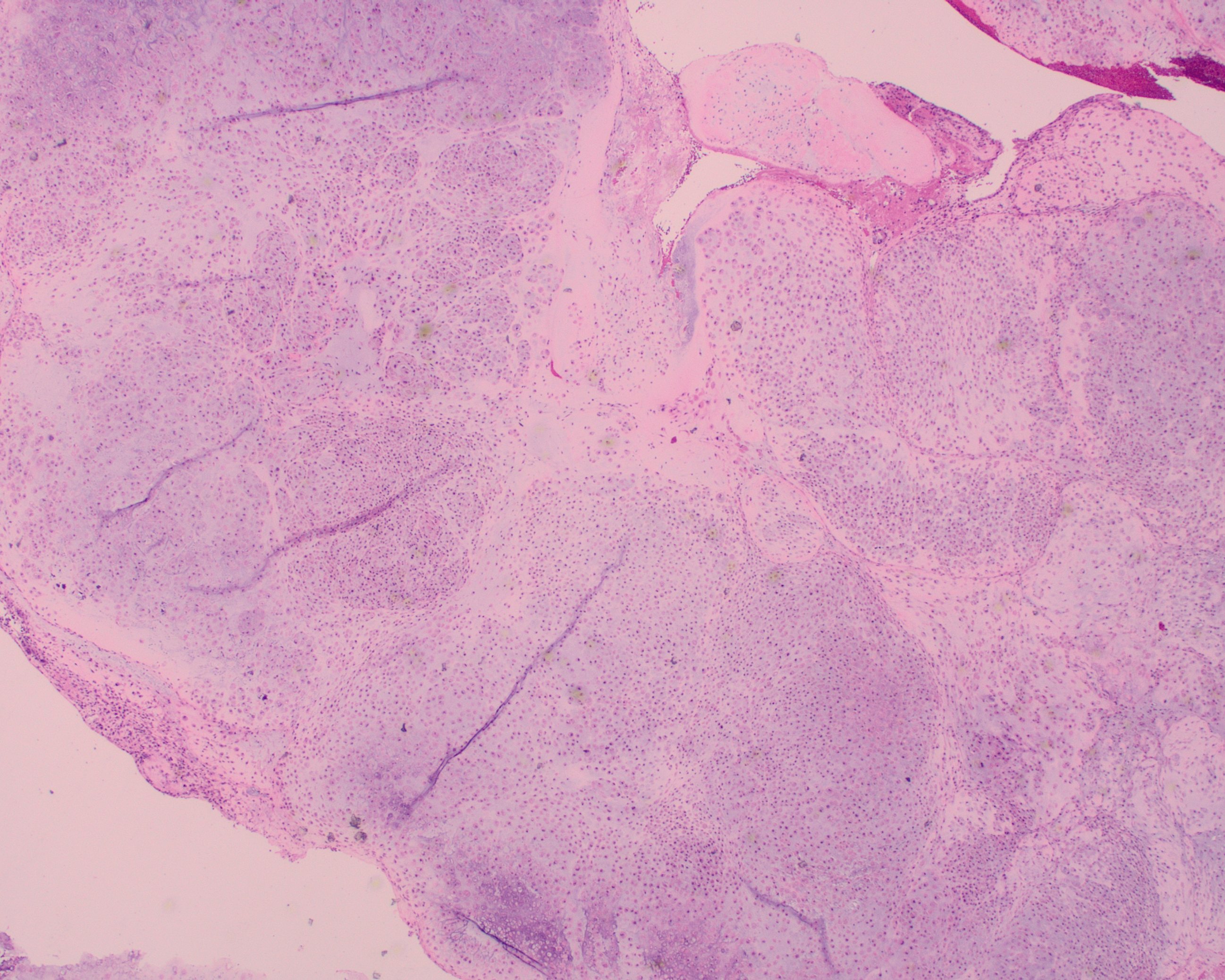
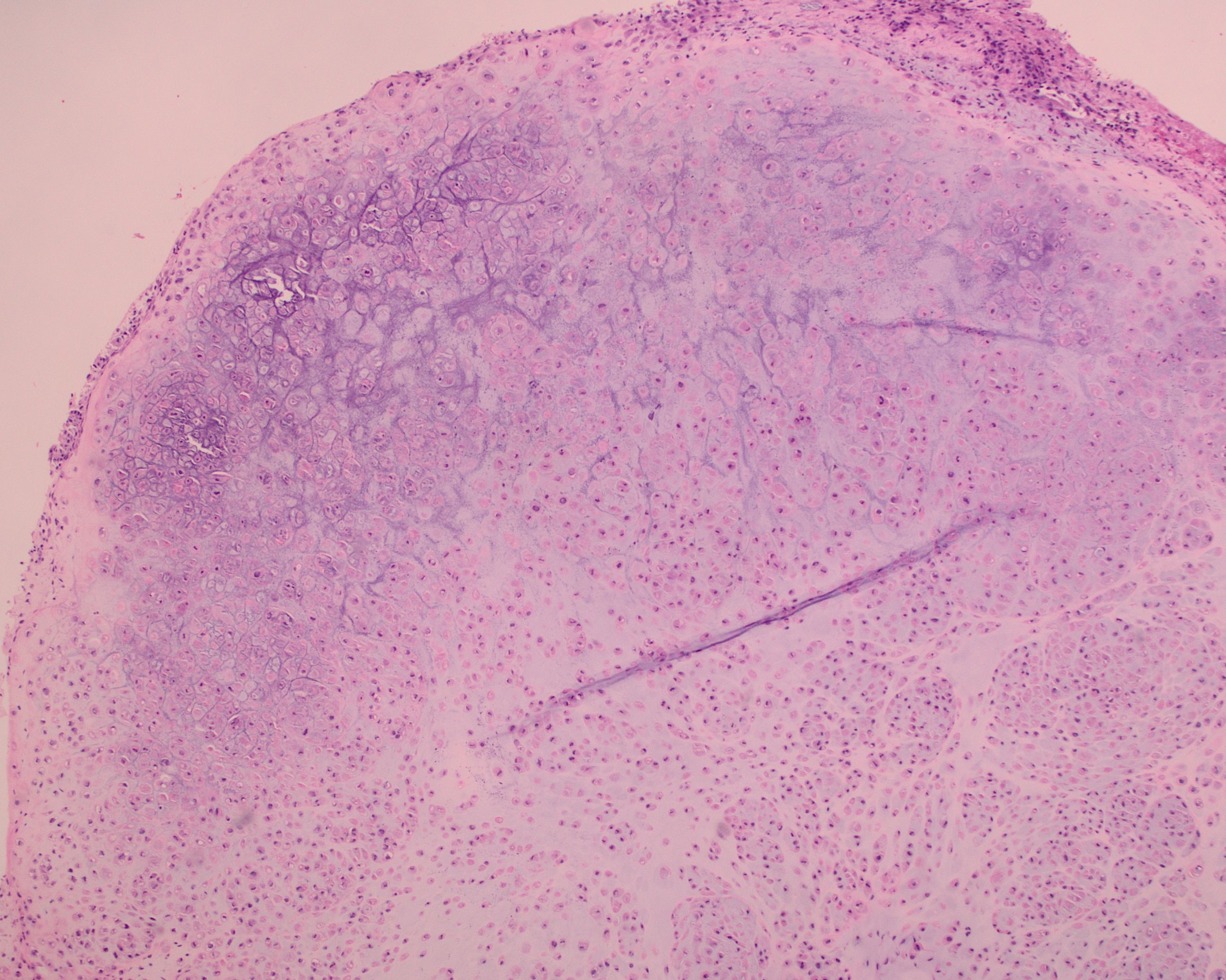
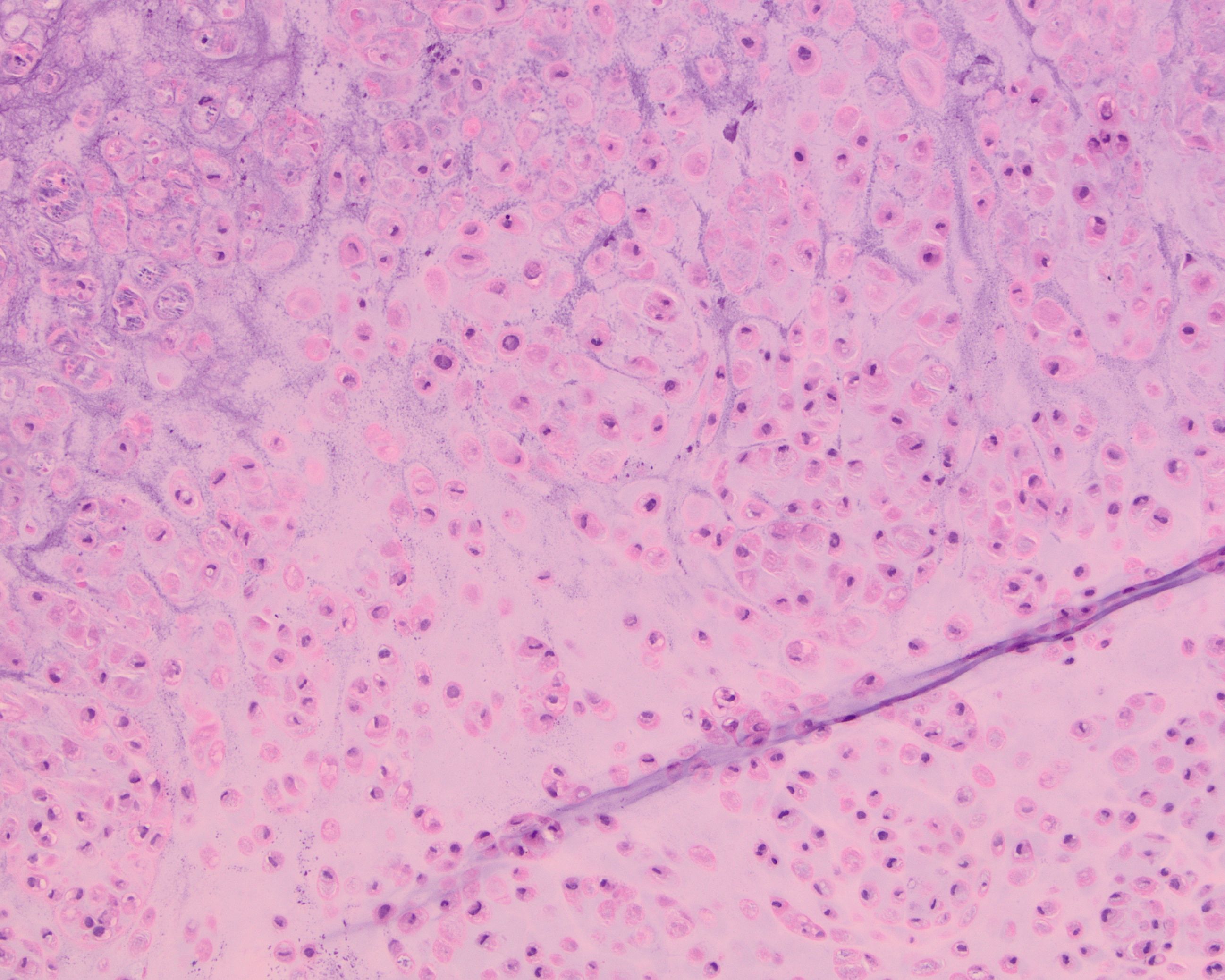
A 55 year-old presents with nodular mass in the temporomandibular joint with erosion of the glenoid fossa. Histologic sections from this lesion are shown below.
What is the most likely diagnosis?
A. Chondroblastoma
B. Synovial chondromatosis
C. Calcified chondroid mesenchymal neoplasm
D. Phosphaturic mesenchymal tumor
Correct Answer: C. Calcified chondroid mesenchymal neoplasm
Discussion:
Next generation sequencing of this tumor identified a FN1::TEK gene fusion. Fusions of FN1 with various receptor tyrosine kinase genes (including TEK, FGFR2, FGFR1, MERTK and NTRK1 have been observed in calcified chondroid mesenchymal neoplasms.
This is a newly recognized group of benign soft‑tissue tumors termed calcified chondroid mesenchymal neoplasms (CCMN). These tumors most often arise in the temporomandibular joint and distal extremities, show nodular/lobular growth of polygonal to stellate cells in a chondroid matrix, and frequently contain grungy or lace‑like (chondroblastoma‑like) calcifications, sometimes with CPPD crystal deposition and focal tenosynovial giant cell tumor (TGCT)-like features. Using targeted RNA sequencing, these tumors harbor recurrent FN1-receptor tyrosine kinase gene fusions in most cases, most commonly FN1::FGFR2, but also FN1::FGFR1 and novel fusions involving MERTK, NTRK1, and TEK, supporting a shared molecular pathogenesis. Although these lesions overlap morphologically with chondroblastoma‑like soft tissue chondroma, chondroid TGCT, and tumoral pseudogout, their distinctive fusion profile and expanded morphologic spectrum justify recognition as a separate diagnostic category with generally indolent behavior after excision.
Reference:
Liu YJ, Wang W, Yeh J, et al. Calcified chondroid mesenchymal neoplasms with FN1-receptor tyrosine kinase gene fusions including FGFR2, FGFR1, MERTK, NTRK1, and TEK: a molecular and clinicopathologic analysis. Mod Pathol. 2021 Jul;34(7):1373-1383.
Case contributed by: Michel Kmeid, M.D., Assistant Professor, Anatomic Pathology, Medical Director, Gross Room, Frozen Section