 |
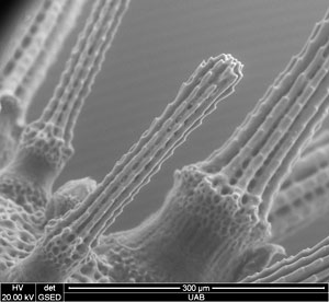
| Images of baby sea urchin. Used environmental SEM mode – image acquired wet. Image acquired for undergraduate biology course. |
When someone says microscope, the first thing that usually comes to mind is those little desktop-sized contraptions used in high school — the ones that magnify a one-inch slide about 500 times its actual size.
Well, move over, Tiny.
The new Scanning Electron Microscope, or SEM, recently purchased by UAB with a $550,000 grant from the National Science Foundation Major Research Instrumentation Program, magnifies images up to 100,000x, and these images can be viewed on the wall of a classroom. The microscope is so powerful that one grain of pollen will look the size of a golf ball, and a human hair, which is roughly 0.0004 inches in diameter, will appear 30 feet across at 100,000x magnification.
Derrick Dean, Ph.D., associate professor of engineering, is the principal investigator on the award. Listed as directing the award are Andrei Stanishevsky, Ph.D., associate professor of physics; Ho-Wook Jun, Ph.D., associate professor of biomedical engineering; Robin Foley, Ph.D., director of the SEM Lab; and Yogesh Vohra, director and associate dean for Interdisciplinary and Creative Innovation in the College of Arts and Sciences.
| The new Scanning Electron Microscope, or SEM, housed in the School of Engineering, is a key addition to UAB’s research arsenal. |
The instrument, housed in the School of Engineering, is a key addition to UAB’s research arsenal, which includes the High-Resolution Imaging Facility’s (HRIF) high-end digital microscopes — including confocal laser microscopy and transmission electron microscopy (TEM). Now, researchers have the ability to see what’s on the surface of their samples and not just through it, says Foley.
Looking at the “face of the object”
“Biological researchers are used to looking through things,” Foley says. “They do a slice of tissue and they look through it on the light microscope or the TEM. The SEM looks at the surface of the samples, and the samples don’t have to be in slices. We can look at bumpy samples, and it enables the researcher to look at the face of the object. We’re going to see the features on the surface, not the features on the interior.”
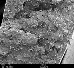 |
| Fractured surface of glass fiber reinforced composite. Image acquired for research in Materials Engineering |
Engineering, Dentistry and the HRIF collaborate to provide support for researchers using the SEM, says Kent Keyser, Ph.D., director of the Vision Science Research Center. Preston Beck, a research associate in prosthodontics, and Melissa Chimento, an electron microscopist in HRIF, provide biological expertise for the SEM.
“The Quanta 650 FEG SEM will have a significant impact on UAB’s research enterprise, in areas ranging from the science of new materials to drug discovery and delivery,” Keyser says. “The availability of state-of-the-art SEM capability will lead to new research collaborations, new cooperative applications for research funding and joint publications. There is a genuine sense of excitement about this new instrument among investigators on campus. School of Engineering faculty who lead this project have done UAB a tremendous service. Speaking on behalf of the staff of the HRIF and the centers that support the facility, I am very excited about working with our colleagues in Engineering and Dentistry to help UAB investigators make the most of this instrument.”
Several centers at UAB provide support for the HRIF, including the Comprehensive Cancer Center, Vision Science Research Center, Hepato/Renal Fibrocystic Disease Core Center, Biomatrix Engineering and Regenerative Medicine (BERM) Center and Comprehensive Arthritis, Musculoskeletal and Autoimmunity Center.
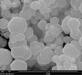 |
| Diamond coatings produced in Department of Physics for Physics Research |
Foley says the new SEM is also part of the UAB Center for Nanoscale Materials and Biointegration (CNMB), led by Vohra. Vohra and Stanishevski, a CNMB professor of physics, played a key role in writing the grant for the SEM. In fact, says Foley, without the backing of the CNMB, UAB might not have been able to get the SEM.
“The new SEM provides a critical characterization tool for many nanoscale science and applications projects. including nanostructured materials for biomedical implants and nanoparticles used in drug delivery and biomedical imaging,” Vohra says.
New advantages
The old microscope was 25 years old when it was retired and only held a one-inch sample, compared to six inches held by the new one. Viewing an object at 10,000x its actual size on the old scope also was a challenge.
“It was an old microscope — older than most of our undergrads by a good bit,” Foley says. “We got a lot of use out of that microscope. It was a star when we bought it, but I was not sad to see it go.”
One advantage of the new microscope is that it’s easy to train students and researchers to use it. Foley had to take as much as six hours to train students to take an image on the old scope. Now, she can have them running their samples and taking pictures in 15 minutes.
| The SEM is available to any investigator or faculty member on campus for use on research projects or for educational purposes. Professors interested in using the SEM to teach or demonstrate to their classes are encouraged to contact Foley and schedule times for their classes to attend. |
“They can do some good, quality research in a short amount of time,” Foley says.
Another advantage is the ability for researchers to image wet materials. The wet materials had to be dried before they could be viewed in the old microscope, and samples sometimes were compromised.
In a recent week, Foley says researchers and students imaged cilia on cells, broken cast iron, tissue and dental implants.
“We can do a wide range of wet or dry samples from 25x to 100,000x magnification,” Foley says. “It’s an extremely wide range.”
Top-end microscope
The Quanta line of scanning electron microscopes are considered versatile, high-performance instruments that can accommodate the widest range of samples of any SEM system. The Quanta 650 FEG is designed with a roomy chamber, enabling the analysis and navigation of large specimens.
The 650 FEG also addresses the need to investigate a variety of materials and characterize structure and composition. Researchers can view almost any sample and obtain surface and compositional images.
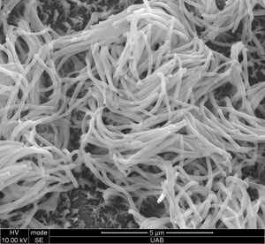 |
| Rabbit septal epithelium. Images acquired for research in Department of Surgery – Otolaryngology |
“We have the top microscope for doing a broad range of samples,” Foley says. “I don’t think there is anything better for doing biological samples. And it can do big samples.”
It also complements very well the confocal microscopes and transmission electron microscopes housed in the HRIF. It gives researchers different tools in their toolbox without having to leave campus.
“We’re a big research institute, and we need all three of these machines — the SEM, TEM and confocal microscopes,” Foley says. “And it’s critical that we work with the other campus microscopy facilities so people don’t waste their time at any location. If I can’t do it, I know where to send them.”
Available to entire campus
The SEM is available to any investigator or faculty member on campus for use on research projects or for educational purposes. Professors interested in using the SEM to teach or demonstrate to their classes are encouraged to contact Foley and schedule times for their classes to attend.
The School of Engineering room where the SEM is set up has television screens for viewing and can accommodate up to 30 students. Graduate students also are welcome to use the microscope by appointment.
The facility also is available for campus tours and for administration or faculty members recruiting perspective employees.
Contact Foley at rfoley@uab.edu or 934-8469 to schedule an appointment.


