 |
Gorgas Case 2019-07 |
|
The following patient was seen in the outpatient clinic of the Infectious Diseases Department of the Hospital Cayetano Heredia during the Gorgas Diploma Course. We would like to thank Dr. Dalila Martinez for her contribution in preparing this case for presentation.
|
 History: A 63-year-old, previously healthy, male patient is admitted with an 8-month history of an indurated erythematous plaque affecting the nose and left maxillary region, rhinorrhea and headache. The patient first reports serous rhinorrhea and nasal congestion eight months prior to admission. Six months prior to admission, he noted an erythematous plaque that began in the left maxillary region. Over the ensuing weeks to months, the plaque progressed despite several rounds of antibiotic therapy and eventually compromised the nose. Due to symptom persistence, the progression of the plaque, the development of nighttime fevers and headaches, the patient was referred to our institution for further evaluation.
Epidemiology: Born and lives in Hualmaca, Chiclayo; a rural area in the northern coast of Peru. Works as a farmer. Frequent contact with cats, dogs, chicken and cattle. Recurrent exposure to fresh-water ponds and rivers. No history of nor exposure to TB. No recent travel history.
Physical examination: Temp: 36.5 °C; HR: 62: RR: 18; BP: 125/76 mmHg; weight: 66.8 kg; O2 Saturation: 97%
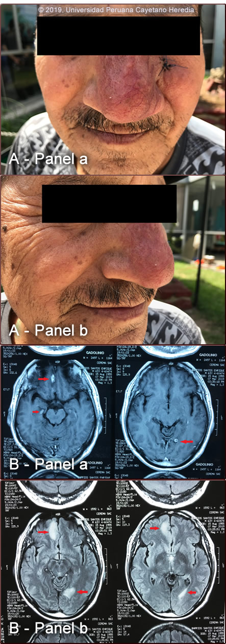
A 7x8 cm infiltrated violaceous plaque with well-defined edges that involves the nose and left maxillary region. There is edema and erythema of the left nares and nasal mucosa [Image A, panels a and b]. No other lesions, discharge or abnormal findings in the oropharynx. Glasgow score of 15, no focal neurologic deficits nor signs of meningeal irritation are found. Muscle strength is preserved symmetrically, bilaterally. The rest of the physical examination is unremarkable. Laboratory result at admission: Hb 10.6 g/dl; MCV: 92; MCH: 30.3; MCHC: 32.8; WBC: 3.4k (N: 61.9 L: 27.8 B: 1 E: 3.6); platelets: 182 000; glucose: 118 mg/dl; urea: 15.4 mg/dl; creatinine: 0.8 mg/dl ; HIV: negative; VDRL: negative.
MRI of the brain: three lesions with diffuse enhancing in the T1-weighted sequence with gadolinium were observed, one in the right frontal lobe and one in the right temporal lobe, and another one in the left occipital lobe [Image B, panel a]. The same lesions looked bright in the FLAIR sequence [Image B, panel b]. UPCH Case Editors: Carlos Seas, Course Director / Carlos McFarlane, Associate Coordinator UAB Case Editors: German Henostroza, Course Director / James Willig, MD / David O. Freedman, Course Director Emeritus |
Diagnosis: Balamuthia mandrillaris (free-living amoeba)
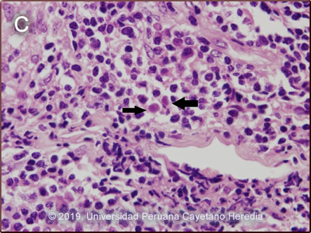
 Discussion: A skin biopsy [Image C] revealed the presence of a lymphoplasmacytic and histiocytic inflammatory infiltrate of moderate intensity with poorly formed granulomas and evidence of multinucleated giant cells. Two structures compatible with amoebic trophozoites were observed. Based on our clinical experience with more than 50 cases and the characteristic skin and brain lesions occurring in concert, a diagnosis of Balamuthia mandrillaris was made. See cases 2007-10, 2004-2, 2015-6 for other patients with these same characteristic manifestations of B. mandrillaris infection. About 200 cases of B. mandrillaris infection have been reported worldwide. Disease has been reported from all continents except Africa. Most cases are reported in the Western hemisphere, with the highest concentration in South America and the United States of America. The disease appears to occur more frequently among patients of Hispanic origin. Possible explanations include genetic susceptibility or high environmental exposure. As in most Latin American cases, our patient was not immunocompromised. Unlike Acanthamoeba and Naegleria species, which are more familiar to clinicians and known to occur in brackish ponds and creeks, an ecologic niche in nature has not been definitively found for Balamuthia. Entry of water into the nasal mucosa and the olfactory nerve endings is thought to precede Naegleria infection. Our patients with Balamuthia infection have come from throughout Peru with most coming from desert areas and not always reporting a history of swimming in potentially contaminated fresh water. Naegleria infection causes an acute necrotizing and suppurative meningoencephalitis, an aggressive disease that is generally fatal in days. Acanthamoeba cause a sub-acute granulomatous encephalitis with a more prolonged but ultimately fatal course mainly seen in immunocompromised hosts. Occasionally, patients with Acanthamoeba may develop a chronic cutaneous ulcer. Acanthamoeba, unlike Balamuthia, has also been associated with amoebic keratitis, a painful sight-threatening disease of the eye which may be associated with contact lens use. In Balamuthia infection, the disease may follow also a prolonged course with an ultimately fatal outcome. Almost all cases have an initial skin lesion preceding CNS disease (granulomatous amebic encephalitis) by weeks or months. The CNS lesions generally appear distant or as metastases from the primary cutaneous lesion. Sometimes as in this case, there may be several metastatic lesions in different parts of the brain and one may seem to be due to direct local extension from the skin lesion [Image B]. Infected patients range from 3 to 65 years of age with 50% under age 15. The typical skin lesion is a single painless plaque up to several centimeters in diameter; a few patients have had 2-3 lesions. Color may be skin tone, dark red, or slightly violaceous. Sensation is preserved. Location is usually on the central face with fewer than 10% with lesions on trunk or extremities. For facial lesions, the differential diagnosis may include tuberculosis, mucocutaneous leishmaniasis, sporotrichosis, leprosy, paracoccidioidomycosis, rhinoscleroma, or atypical mycobacteria. Sarcoid, discoid lupus, cutaneous lymphoma (NK lymphoma) and Wegener’s can also be considered. Histologically, granulomatous inflammation with lymphocytes, histiocytes, plasma cells, as well as giant cells, is characteristic. Amoebic trophozoites are often rare and multiple sections need to be examined for their detection. Some foci of vasculitis may be present as well. Balamuthia CNS involvement most often manifests with headache, photophobia, seizures that progress to lethargy, sensori-motor deficit(s), coma and death. CNS lesions undergo progressive hemorrhagic necrosis with large numbers of amoebic trophozoites and cysts invading vascular structures (sub-adventitial areas of arteries, veins, and capillaries), leading to perivasculitis and cerebral infarcts [Hum Pathol. 1999 Mar;30(3):269-73]. The mainstay of successful treatment for Balamuthia depends on early diagnosis and therapy. Patients without brain lesion(s) at diagnosis have better chances of recovery. Treatment with drug combinations for prolonged periods is warranted. This patient was started on fluconazole, albendazole, and miltefosine based on our prior experience with one patient [Clin Infect Dis. 2010;51(2):e7-11]. Our center has treated 12 patients with and without CNS involvement since that publication with this three-drug combination, 7 survived with complete clearance of the lesions [Martínez DY. Personal communication]. This drug regimen is well tolerated and the most important side effects are nausea, stomach ache, and vomiting. Treatment ranged from 6 to 18 months in most of these cases. Miltefosine crosses the blood-brain barrier and is known to have an in-vitro amebicide effect against B. mandrillaris [J Eukaryot Microbiol. 2006 Mar-Apr;53(2):121-6]. Although formal trials are lacking a number of case reports have shown good results when using miltefosine as part of a combination of drugs to treat free-living amebic infections including Naegleria sp. Acanthamoeba and B. mandrillaris. Miltefosine is FDA-approved and available in the US but therapy is costly and there is a single manufacturer. Fluconazole produces an amebistatic effect by interfering with ergosterol metabolism (ergosterol is one of the main components of the amebic wall). Albendazole affects cell motility, maintenance of cell shape, intracellular transport and diminishes ATP production. Both drugs, have good CNS penetration. Other drugs with amebicidal effects are: pentamidine and some antipsychotic agents (phenothiazines and thioridazine). Unfortunately, they are poorly tolerated. While IV amphotericin B or pentamidine, have produced initial, apparently favorable responses including disappearance of cutaneous lesions, their administration still does not halt the eventual appearance of CNS disease. Similarly, in our experience, multi-drug combination therapy with agents such as albendazole, itraconazole, or fluconazole without miltefosine do not halt the eventual appearance of or progression to CNS disease. |